McKinlay & Peters Equine Hospital
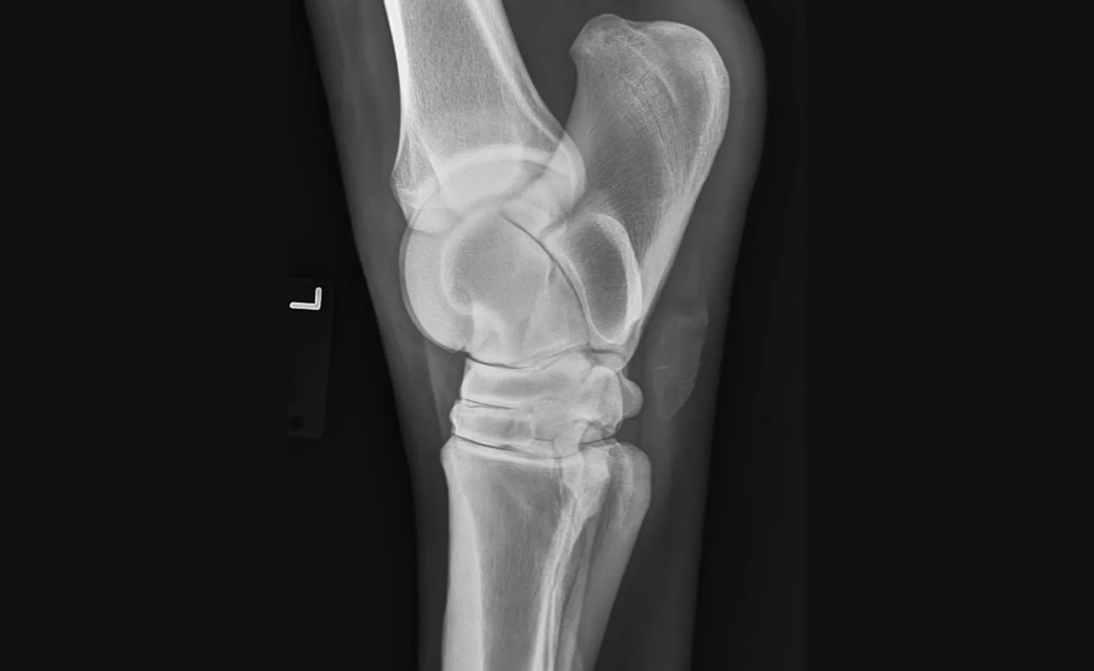
Digital Radiography
Digital radiography allows for evaluation of bones, both normal and abnormal, which can be used in several different types of appointments. Images are high quality and can be viewed immediately to provide timely recommendations. Wounds and (some) foreign objects can be evaluated and located. Intra-operative radiographs ensure proper placement of orthopedic screws and plates.
Arthritis and lameness
Dentistry
Fractures
Joint injections
Surgery
Wound evaluation
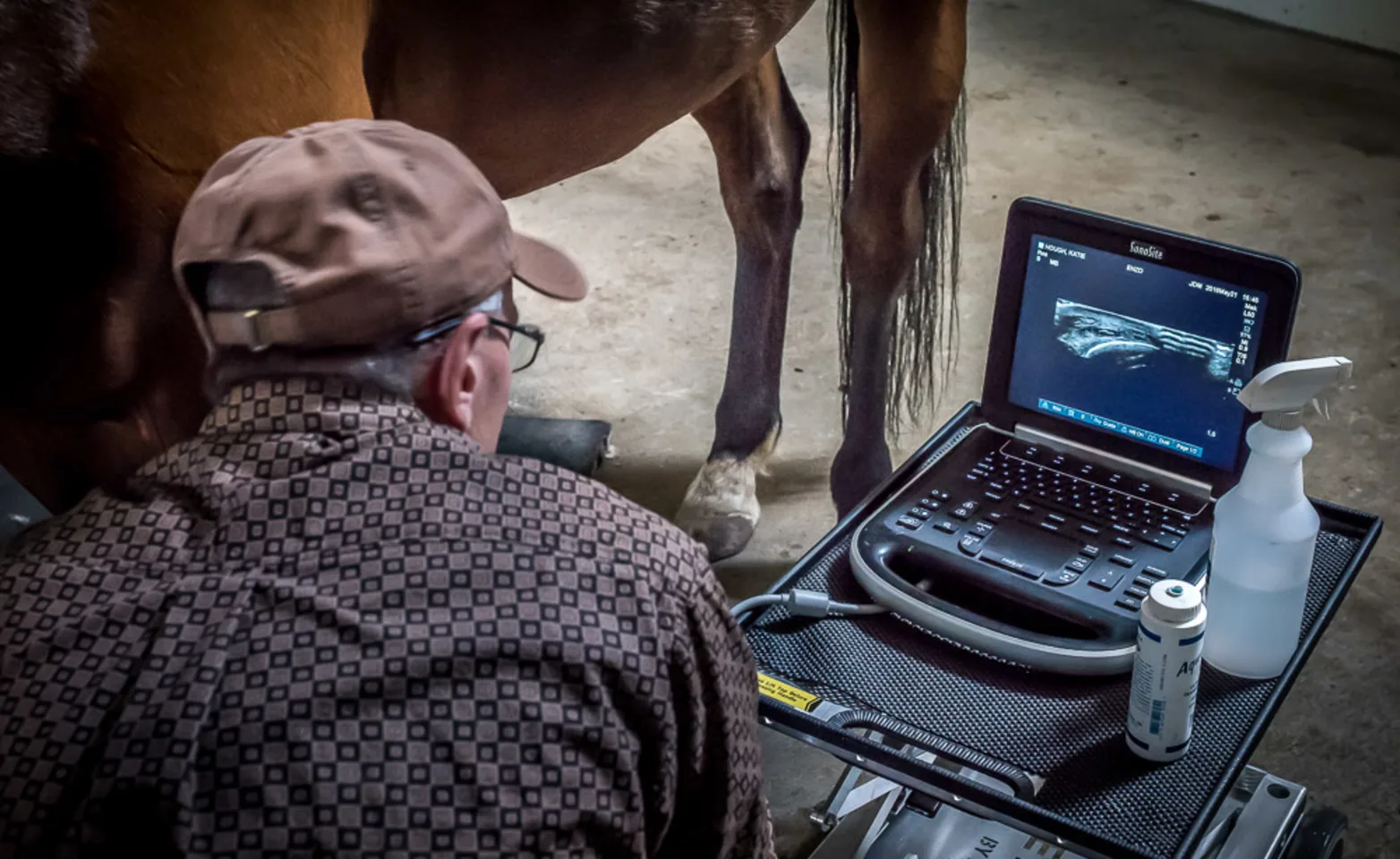
Ultrasonography
Ultrasonography utilizes high-frequency sounds waves for non-invasive examination of soft tissue structures and internal organs. Like radiography, it is used in several different appointments, particularly reproduction. Ultrasound allows for real-time evaluation, which provides information to make immediate recommendations.
Colic
Joint injections
Lameness
Pneumonia
Mass examination
Reproduction
Wound evaluation
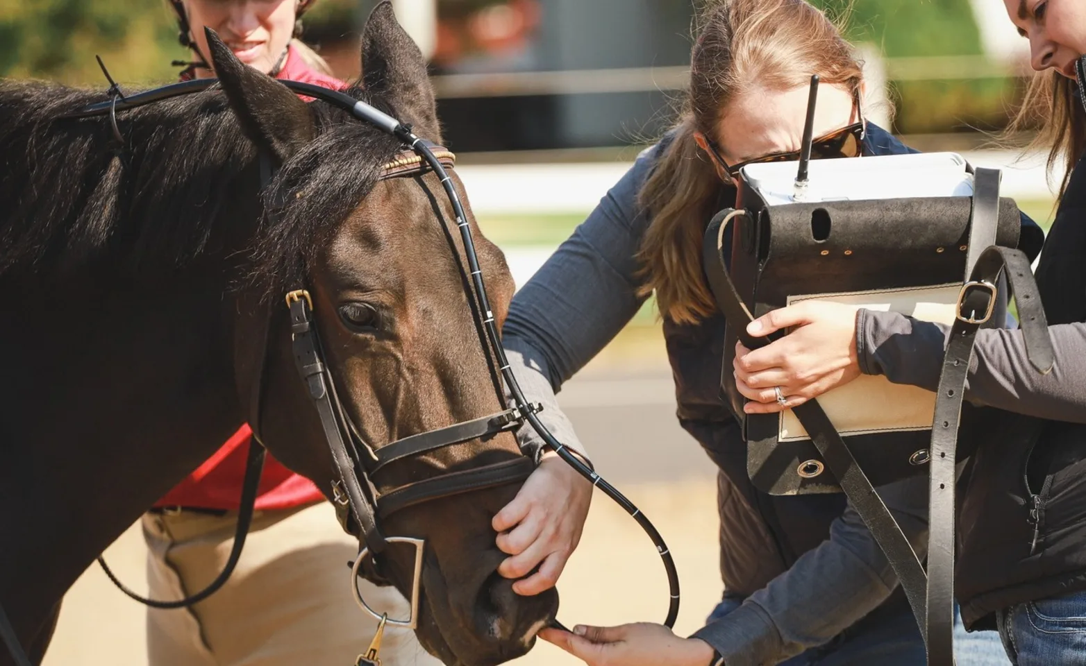
Dynamic upper airway endoscopy
Several variables (tack type/placement, head position, speed, etc.) affect respiration while working. Static endoscopy often requires sedation, making it difficult to evaluate the effect these variables have on your horse. Dynamic endoscopy involves real-time monitoring at various levels of work. While most often used to evaluate the inspiration/expiration processes, dynamic endoscopy can also help diagnose issues while swallowing.
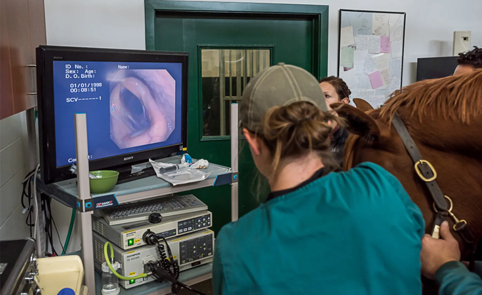
Endoscopy
Endoscopic procedures involve passing a long camera into a body cavity or opening, allowing us to visualize internal structures. MPEH has two lengths of flexible scopes, allowing for evaluation of both ends of your horse. Diagnosis of stomach ulcers and bladder abnormalities, as well as respiratory and gastrointestinal issues, is improved when using flexible endoscopy. In addition, MPEH has a fixed scope for joint and minimally invasive abdominal surgeries.
Arthroscopy (joint)
Colonoscopy (large intestine)
Cystoscopy (urinary bladder)
Esophagoscopy (esophagus)
Gastroscopy (stomach)
Laparoscopy (abdominal)
Pharyngoscopy (upper respiratory tract)
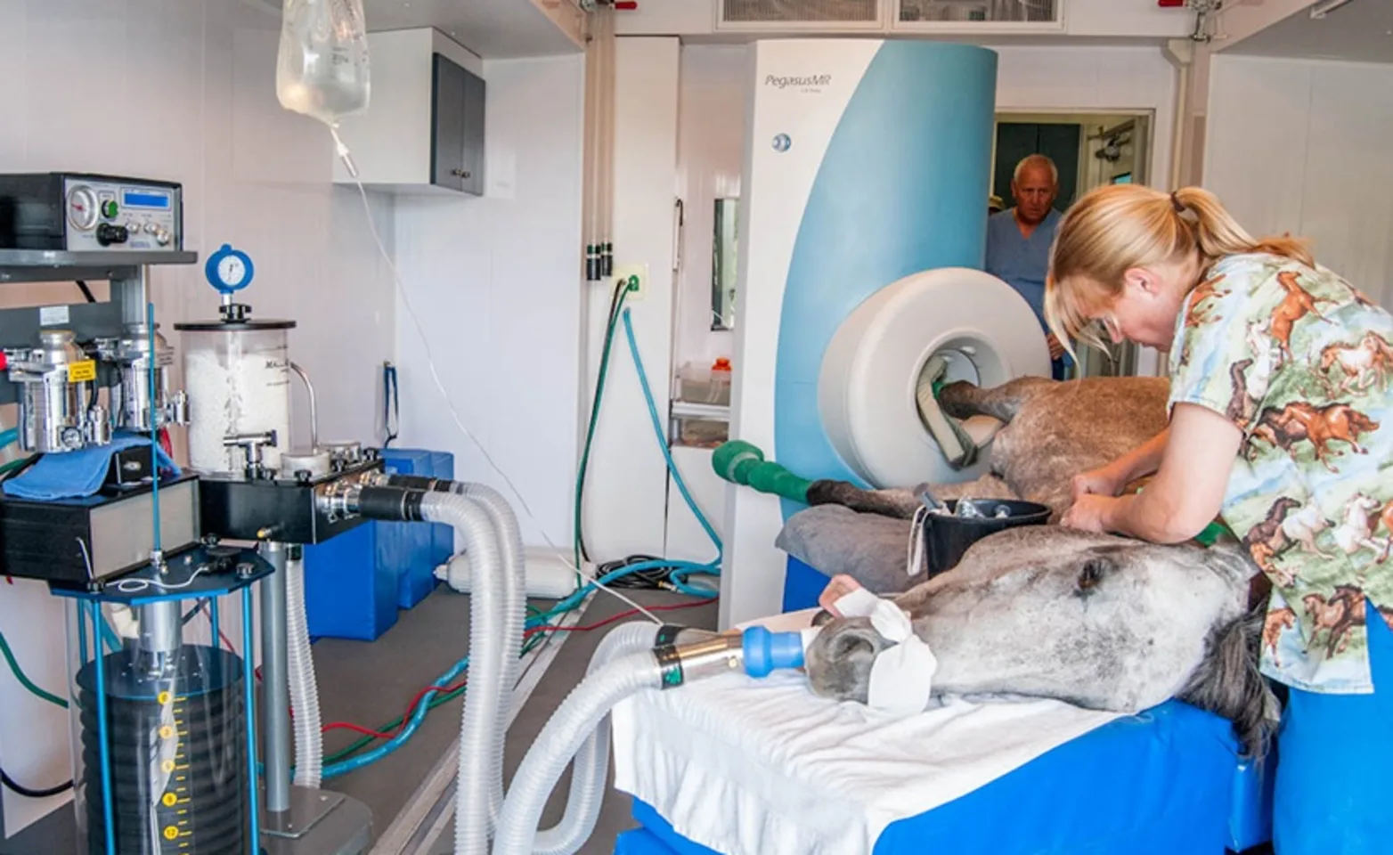
Equine MRI – Newman Lake Hospital
The number one thing a veterinarian needs to know when treating a horse with a lameness – is the cause. The guesswork is eliminated when the problem is accurately identified. This greatly improves the horse’s chances of returning to performance. Precise diagnosis leads to proper treatment, which leads to the best possible prognosis. Magnetic Resonance Imaging (MRI) is a powerful diagnostic tool that allows us to see bone, cartilage, tendon, & ligaments. MRI studies allow for the highest level of diagnostic accuracy. If your horse has a lameness problem that cannot be diagnosed, you should strongly consider having an MRI study performed. It is imperative to keep in mind that not all MRI evaluations are the same. High-field magnets (1.0 or 1.5 Tesla) provide unsurpassed accuracy & detail when compared to low-field or standing magnets. Often the problem can be overlooked when studies are performed with low-field magnets.
